SOLDES
Jusqu'à -70% sur une sélection d'articles*

A Simple Guide to Venous Ulcers, Diagnosis, Treatment and Related Conditions
Par :Formats :
Disponible dans votre compte client Decitre ou Furet du Nord dès validation de votre commande. Le format ePub est :
- Compatible avec une lecture sur My Vivlio (smartphone, tablette, ordinateur)
- Compatible avec une lecture sur liseuses Vivlio
- Pour les liseuses autres que Vivlio, vous devez utiliser le logiciel Adobe Digital Edition. Non compatible avec la lecture sur les liseuses Kindle, Remarkable et Sony
 , qui est-ce ?
Notre partenaire de plateforme de lecture numérique où vous retrouverez l'ensemble de vos ebooks gratuitement
Pour en savoir plus sur nos ebooks, consultez notre aide en ligne ici
C'est si simple ! Lisez votre ebook avec l'app Vivlio sur votre tablette, mobile ou ordinateur :

- FormatePub
- ISBN978-1-005-39554-4
- EAN9781005395544
- Date de parution26/11/2021
- Protection num.pas de protection
- Infos supplémentairesepub
- ÉditeurC. C. Chamberlane
Résumé
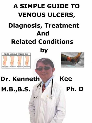
This book describes Venous Ulcers, Diagnosis and Treatment and Related DiseasesThe worst venous ulcers that I have seen are those from diabetes and varicose veins. Venous ulcers (also termed venous stasis ulcers or non-healing wounds) are open wounds happening around the ankle or lower leg. They do not recover for weeks or months, and occasionally continue for longer. Venous ulcers (VUs) are late markers of chronic venous insufficiency (CVI) and venous hypertension.
In normal disorders, calf muscle contraction and intra-luminal valves stimulate prograde flow while stopping blood reflux. When retrograde flow, obstruction, or both are present, the resultant chronic venous hypertension is accountable for the dermatologic and vascular complications that result in the formation of a VU. Some disorders that can cause venous ulcers are:Venous hypertensionVenous insufficiencyVaricose veinsBlood clots or poor circulationDiabetes, kidney failure, or inflammatory diseasesCertain medicinesInfectionsObesityA venous leg ulcer can form after a minor injury if there is a disorder with the circulation of blood in the leg veins.
When this happens, the pressure within the veins rises. This constant high pressure can slowly injure the tiny blood vessels in the skin and make it fragile. As a result, the skin can easily tear and form an ulcer after a hit or scratch. Unless the patient has treatment to increase the circulation in the legs, the ulcer may not recover. Risk factorsA number of factors can raise the risk of developing a venous leg ulcer, such as:Obesity or being overweight - raises the pressure in the leg veinsDifficulty in walking - weaken the calf muscles, which can affect circulation in the leg veinsPrevious deep vein thrombosis (DVT) - blood clots in the leg can injure the valves in the veinsVaricose veins - swollen and enlarged veins produced by malfunctioning valvesPrevious injury to the leg, such as a broken or fractured bone, which may produce DVT or impair walkingPrevious surgery to the leg, such as a hip replacement or knee replacement, which can prevent the patient movingAge - people find it harder to move around as they ageWhile CVI is a well-known trigger for VU development, venous ulceration happens rarely (5.1%) for unknown reasons.
CVI may happen due to blood reflux, obstruction, or a combination of both mechanisms, producing macro- and micro-circulatory dysfunction. If the patient has a venous leg ulcer, the patient may also have symptoms:Swollen anklesDiscoloration and darkening of the skin around the ulcerHardened skin around the ulcer, which may make the leg feel hardHeavy feeling in the legsAching or swelling in the legsRed, flaky, scaly and itchy skin on the legsColor-flow duplex ultrasound is another non-costly, non-invasive, and highly informative diagnostic test, especially useful for superficial vein assessment.
The method can identify thrombi presence and valve incompetence, which is affirmed when reflux time is > 0.5 s. Computed tomography and magnetic resonance imaging are favored for deeper vessels since they are often more difficult or impossible to evaluateThe main treatment for VUs depends on two methods: compression therapy and direct wound management. Occasionally minor surgery (wound debridement) or vein ablation is used to remove any nonliving tissue that stops a wound from healing properly.
With proper treatment, most venous leg ulcers recover within 3 - 4 months. TABLE OF CONTENTIntroductionChapter 1 Venous UlcersChapter 2 CausesChapter 3 SymptomsChapter 4 DiagnosisChapter 5 TreatmentChapter 6 PrognosisChapter 7 Varicose Veins...
In normal disorders, calf muscle contraction and intra-luminal valves stimulate prograde flow while stopping blood reflux. When retrograde flow, obstruction, or both are present, the resultant chronic venous hypertension is accountable for the dermatologic and vascular complications that result in the formation of a VU. Some disorders that can cause venous ulcers are:Venous hypertensionVenous insufficiencyVaricose veinsBlood clots or poor circulationDiabetes, kidney failure, or inflammatory diseasesCertain medicinesInfectionsObesityA venous leg ulcer can form after a minor injury if there is a disorder with the circulation of blood in the leg veins.
When this happens, the pressure within the veins rises. This constant high pressure can slowly injure the tiny blood vessels in the skin and make it fragile. As a result, the skin can easily tear and form an ulcer after a hit or scratch. Unless the patient has treatment to increase the circulation in the legs, the ulcer may not recover. Risk factorsA number of factors can raise the risk of developing a venous leg ulcer, such as:Obesity or being overweight - raises the pressure in the leg veinsDifficulty in walking - weaken the calf muscles, which can affect circulation in the leg veinsPrevious deep vein thrombosis (DVT) - blood clots in the leg can injure the valves in the veinsVaricose veins - swollen and enlarged veins produced by malfunctioning valvesPrevious injury to the leg, such as a broken or fractured bone, which may produce DVT or impair walkingPrevious surgery to the leg, such as a hip replacement or knee replacement, which can prevent the patient movingAge - people find it harder to move around as they ageWhile CVI is a well-known trigger for VU development, venous ulceration happens rarely (5.1%) for unknown reasons.
CVI may happen due to blood reflux, obstruction, or a combination of both mechanisms, producing macro- and micro-circulatory dysfunction. If the patient has a venous leg ulcer, the patient may also have symptoms:Swollen anklesDiscoloration and darkening of the skin around the ulcerHardened skin around the ulcer, which may make the leg feel hardHeavy feeling in the legsAching or swelling in the legsRed, flaky, scaly and itchy skin on the legsColor-flow duplex ultrasound is another non-costly, non-invasive, and highly informative diagnostic test, especially useful for superficial vein assessment.
The method can identify thrombi presence and valve incompetence, which is affirmed when reflux time is > 0.5 s. Computed tomography and magnetic resonance imaging are favored for deeper vessels since they are often more difficult or impossible to evaluateThe main treatment for VUs depends on two methods: compression therapy and direct wound management. Occasionally minor surgery (wound debridement) or vein ablation is used to remove any nonliving tissue that stops a wound from healing properly.
With proper treatment, most venous leg ulcers recover within 3 - 4 months. TABLE OF CONTENTIntroductionChapter 1 Venous UlcersChapter 2 CausesChapter 3 SymptomsChapter 4 DiagnosisChapter 5 TreatmentChapter 6 PrognosisChapter 7 Varicose Veins...
A propos de Kenneth Kee


4,49 €