

The Craniotomy Atlas
Par :Formats :
- Paiement en ligne :
- Livraison à domicile ou en point Mondial Relay indisponible
- Retrait Click and Collect en magasin gratuit
- Nombre de pages237
- PrésentationRelié
- FormatGrand Format
- Poids1.315 kg
- Dimensions23,0 cm × 30,0 cm × 2,0 cm
- ISBN978-3-13-205791-3
- EAN9783132057913
- Date de parution10/07/2019
- ÉditeurThieme
- Directeur de publicationBernhard Meyer
- Directeur de publicationKarl Schaller
- Directeur de publicationPeter Vajkoczy
- Directeur de publicationPeter Winkler
Résumé
Given that the great majority of brain surgeries are preceded by a craniotomy, mastering the procedure is essential for junior residents. Choosing the appropriate craniotomy and executing it safely is the difference between a straightforward case with good access to the target and a procedure where access to the target is needlessly traumatic and may even be impossible. Professor Raabe's "The Craniotomy Atlas" provides precise instructions for performing all common neurosurgical cranial exposures, including : convexity approaches, midline approaches, skull base approaches, transsphenoidal approaches, and more.
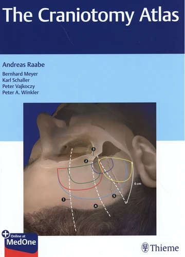
Instructions for each craniotomy include positioning, head fixation, aesthetic considerations. and protecting the dura mater. Special Features : More than 900 high-quality operative photographs and brilliant illustrations support the step-by-step descriptions, with all the precision and attention to detail that neurosurgeons have come to expect from the editor, Professor Raabe, and the associate editors, Professors Meyer, Schaller, Vajkoczy, and Winkler ; Full coverage of complications and risk factors ; Checklist with summaries of the critical steps.
All residents and trainees in neurosurgery will treasure this essential resource, which will help build confidence when performing these critical neurosurgical procedures.
Given that the great majority of brain surgeries are preceded by a craniotomy, mastering the procedure is essential for junior residents. Choosing the appropriate craniotomy and executing it safely is the difference between a straightforward case with good access to the target and a procedure where access to the target is needlessly traumatic and may even be impossible. Professor Raabe's "The Craniotomy Atlas" provides precise instructions for performing all common neurosurgical cranial exposures, including : convexity approaches, midline approaches, skull base approaches, transsphenoidal approaches, and more.
Instructions for each craniotomy include positioning, head fixation, aesthetic considerations. and protecting the dura mater. Special Features : More than 900 high-quality operative photographs and brilliant illustrations support the step-by-step descriptions, with all the precision and attention to detail that neurosurgeons have come to expect from the editor, Professor Raabe, and the associate editors, Professors Meyer, Schaller, Vajkoczy, and Winkler ; Full coverage of complications and risk factors ; Checklist with summaries of the critical steps.
All residents and trainees in neurosurgery will treasure this essential resource, which will help build confidence when performing these critical neurosurgical procedures.