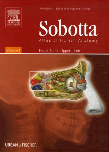
Sobotta Atlas of Human Anatomy. Tome 1, Head, Neck, Upper Limb
14e édition
Par : , Formats :
- Paiement en ligne :
- Livraison à domicile ou en point Mondial Relay indisponible
- Retrait Click and Collect en magasin gratuit
- Réservation en ligne avec paiement en magasin :
- Indisponible pour réserver et payer en magasin
- Nombre de pages419
- PrésentationRelié
- Poids2.265 kg
- Dimensions23,5 cm × 32,0 cm × 3,0 cm
- ISBN0-443-10348-8
- EAN9780443103483
- Date de parution01/04/2006
- ÉditeurChurchill Livingstone
- TraducteurS Bedoui
Résumé
The complete macroscopic anatomy. The two-volume Sobotta Atlas covers the complete macroscopic anatomy in full detail and unequalled quality with almost 2000 figures. The 14th edition contains more than 200 new figures. Easy to manage: As in the dissection course, the Atlas is organised by body regions - this corresponds to the topics for course attendance certificates. Simple new introductory schemes and general overviews help you to understand the more complex figures and connections - step by step. Learning for clinical practice from the beginning: To facilitate the application of anatomical knowledge to clinical practice, the new Sobotta comes with even more clinical figures, including new imaging techniques (X-ray, MRI, CT etc.), endoscopic images, intraoperative colour photographs of inner organs, pictures from patients with common paralyses and more - designed to be suitable for any type of curriculum. Especially handy: Volume 1 is accompanied by a compact brochure with tables of muscles, joints, and nerves.
The complete macroscopic anatomy. The two-volume Sobotta Atlas covers the complete macroscopic anatomy in full detail and unequalled quality with almost 2000 figures. The 14th edition contains more than 200 new figures. Easy to manage: As in the dissection course, the Atlas is organised by body regions - this corresponds to the topics for course attendance certificates. Simple new introductory schemes and general overviews help you to understand the more complex figures and connections - step by step. Learning for clinical practice from the beginning: To facilitate the application of anatomical knowledge to clinical practice, the new Sobotta comes with even more clinical figures, including new imaging techniques (X-ray, MRI, CT etc.), endoscopic images, intraoperative colour photographs of inner organs, pictures from patients with common paralyses and more - designed to be suitable for any type of curriculum. Especially handy: Volume 1 is accompanied by a compact brochure with tables of muscles, joints, and nerves.


